The de facto standard of resolution evaluation charts for X-ray analysis.
Benefits
NTT-AT's X-ray resolution evaluation charts are applied to several X-ray analysis situations which require ultra-high resolution, such as X-ray microscopes, X-ray micro-beam analysis, and X-ray imaging. This de facto standard X-ray chart is used in a very large number of situations throughout the world.
High X-ray irradiation durability, ultra-sharp pattern, and low edge roughness. These are the biggest features of NTT-AT's X-ray chart. Our SiC membrane based Ta absorber chart has proved to be outstandingly accurate and provides clear images for your X-ray analysis system evaluation.
Please try out the proven performance as the de facto standard.Features
Three types of X-ray chart are available for various applications: standard type, high resolution and high contrast type, and ultra-high resolution type. Customization of the pattern layout and substrate dimensions is available for your system.Specifications
| Item | Standard type
XRESO-100 | High resolution type
with thicker Ta
absorber
XRESO-50HC | NEW!!
Ultra high resolution
XRESO-20 |
|---|
| Substrate | Material / Size | Si 10mm square |
|---|
| Thickness | 1mm | 1mm | 0.625mm |
| Membrane | Material /
Thickness | Ru 20nm
SiN 2µm | Ru 20nm
SiC 200nm
SiN 50nm | Ru 20nm
SiC 200nm
SiN 50nm |
|---|
| Area | 1mm square | 1mm square | 1mm square |
| Alignment | Center of the substrate | Center of the substrate | Center of the substrate |
| Pattern | Absorber /
Thickness | Ta 1µm | Ta 500nm | Ta 100nm |
|---|
Minimum pattern
size | 100nm | 50nm | 20nm
Radial Pattern |
| Patterned area | 250µm × 350µm | 300µm square | 300µm square |

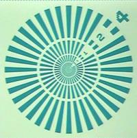
Schematic of X-ray chart (High resolution chart)
X-ray chart Inquiry
Ultra-high resolution type
XRESO-20
XRESO-20 is the ultra-high resolution evaluation chart featuring minimum patern width of 20nm . This high specification model, released for sale in 2014, is applied to recent ultra-high resolution X-ray imaging system.
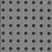
| SEM image of chart | Pattern layout |
|---|
| 100nm hole | 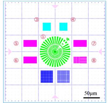
①Radial Pattern
③④Hole Pattern
⑤⑥⑦⑧L&S Pattern |
 |
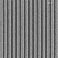
| 50nm L&S |
 |
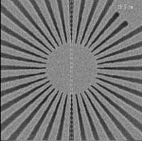
| 20nm patterns | 20nm Radial patterns |
 |  |
X-ray chart Inquiry
Standard type
XRESO-100
XRESO-100 provides a resolution of 100nm at low cost. This high contrast standard chart has not only been applied to scientific fields but also to industry use, such as the calibration of X-ray inspection system.| Pattern layout |
|---|
 |
X-ray chart Inquiry
High resolution type with thicker absorber
XRESO-50HC
XRESO-50HC provides a high resolution of 50nm at a reasonable cost. It has been applied to various applications such as X-ray micro beam irradiation, X-ray microscopes, and X-ray coherent imaging.| SEM image of chart | Pattern layout |
|---|
Radial patterns
Corresponding to point (1) of the pattern layout |  |
 |
50nm L&S
Corresponding to point (2) of the pattern layout |
 |
X-ray chart Inquiry
Past record
NTT-AT's de facto standard X-ray resolution evaluation charts are utilized in worldwide enterprises, universities, and research institutes.
Paper lists
- Satoshi Matsuyama, Yoji Emi, Hidetoshi Kino, Yoshiki Kohmura, Makina Yabashi, Tetsuya Ishikawa, and Kazuto Yamauchi, “Achromatic and high-resolution full-field X-ray microscopy based on total-reflection mirrors,” Opt. Express 23, 9746 (2015); http://dx.doi.org/10.1364/OE.23.009746
- Shinji Ohsuka, Akira Ohba, Shinobu Onoda, Katsuhiro Nakamoto, Tomoyasu Nakano, Motosuke Miyoshi, Keita Soda and Takao Hamakubo, “Laboratory-size three-dimensional X-ray microscope with Wolter type I mirror optics and an electron-impact water window X-ray source ,” Rev. Sci. Instrum. 85, 093701 (2014); http://dx.doi.org/10.1063/1.4894468
- A. Schropp, P. Boye, J. M. Feldkamp, R. Hoppe, J. Patommel, D. Samberg, S. Stephan, K. Giewekemeyer, R. N. Wilke, T. Salditt, J. Gulden, A. P. Mancuso, I. A. Vartanyants, E. Weckert, S. Schöder, M. Burghammer and C. G. Schroer, “Hard X-ray nanobeam characterization by coherent diffraction microscopy,” Appl. Phys. Lett. 96, 091102 (2010); http://dx.doi.org/10.1063/1.3332591
- A. Schropp, R. Hoppe, J. Patommel, D. Samberg, F. Seiboth, S. Stephan, G. Wellenreuther, G. Falkenberg and C. G. Schroer, “Hard X-ray scanning microscopy with coherent radiation: Beyond the resolution of conventional X-ray microscopes,” Appl. Phys. Lett. 100, 253112 (2012); http://dx.doi.org/10.1063/1.4729942
- P. Bruyndonckxa, A. Sasova and B. Pauwelsa, “Towards sub-100-nm X-ray microscopy for tomographic applications,” Powder Diffr. 25, 157 (2010); http://dx.doi.org/10.1154/1.3416936
- S. P. Krüger, K. Giewekemeyer, S. Kalbfleisch, M. Bartels, H. Neubauer, and T. Salditt, “Sub-15 nm beam confinement by two crossed X-ray waveguides,” Opt. Express 18, 13492 (2010); http://dx.doi.org/10.1364/OE.18.013492
- J. W. Jung, J. S. Lee, N. Kwon, S. J. Park, S. Chang, J. Kim, J. Pyo, Y. Kohmura, Y. Nishino, M. Yamamoto, T. Ishikawa and J. H. Je, “Fast microtomography using bright monochromatic X-rays,” Rev. Sci. Instrum. 83, 093704 (2012); http://dx.doi.org/10.1063/1.4751853
- Sven Niese, Peter Krüger, Adam Kubec, Stefan Braun, Jens Patommel, Christian G. Schroer, Andreas Leson, and Ehrenfried Zschech, Full-field X-ray microscopy with crossed partial multilayer Laue lenses,” Opt. Express 22, 20008 (2014); http://dx.doi.org/10.1364/OE.22.020008
- Mike Beckers, Tobias Senkbeil, Thomas Gorniak, Klaus Giewekemeyer, Tim Salditt and Axel Rosenhahn, “Drift correction in ptychographic diffractive imaging,” Ultramicroscopy 126, 44 (2013); http://dx.doi.org/10.1016/j.ultramic.2012.11.006
- Akihiro Suzuki, Shin Furutaku, Kei Shimomura, Kazuto Yamauchi, Yoshiki Kohmura, Tetsuya Ishikawa, and Yukio Takahashi, “High-Resolution Multislice X-Ray Ptychography of Extended Thick Objects,” Phys. Rev. Lett. 112, 053903 (2014); http://dx.doi.org/10.1103/PhysRevLett.112.053903
- Yoshio Suzuki , “Interaction between periodic structures of object and X-ray standing wave generated by wavefront-division interferometer,” Rev. Sci. Instrum. 86, 043701 (2015); http://dx.doi.org/10.1063/1.4916735
- Matthias Müller, Tobias Mey, Jürgen Niemeyer, and Klaus Mann, “Table-top soft X-ray microscope using laser-induced plasma from a pulsed gas jet,” Opt. Express 19, 23489 (2014); http://dx.doi.org/10.1364/OE.22.023489
- F. Seiboth, M. Scholz, J. Patommel, R. Hoppe, F. Wittwer, J. Reinhardt, J. Seidel, M. Knaut, A. Jahn, K. Richter, J. W. Bartha, G. Falkenberg and C. G. Schroer, “Hard X-ray nanofocusing by refractive lenses of constant thickness,” Appl. Phys. Lett. 105, 131110 (2014); http://dx.doi.org/10.1063/1.4896914
- R. N. Wilke, M. Vassholz and T. Salditt, “Semi-transparent central stop in high-resolution X-ray ptychography using Kirkpatrick–Baez focusing,” Acta Cryst. A 69, 490 (2013); http://dx.doi.org/10.1107/S0108767313019612
- T. Salditt, S. Kalbfleisch, M. Osterhoff, S. P. Krüger, M. Bartels, K. Giewekemeyer, H. Neubauer, and M. Sprung, “Partially coherent nano-focused X-ray radiation characterized by Talbot interferometr,” Opt. Express 19, 9656 (2011); http://dx.doi.org/10.1364/OE.19.009656
- K. Giewekemeyer, M. Beckers, T. Gorniak, M. Grunze, T. Salditt, and A. Rosenhahn. “Ptychographic coherent X-ray diffractive imaging in the water window,” Opt. Express 19 1037 (2011); http://dx.doi.org/10.1364/OE.19.001037
- C. Homann, T. Hohage, J. Hagemann, A.-L. Robisch, and T. Salditt, “Validity of the empty-beam correction in near-field imaging,” Phys. Rev. A 91, 013821 (2015); http://dx.doi.org/10.1103/PhysRevA.91.013821
- Klaus Giewekemeyer, Hugh T. Philipp, Robin N. Wilke, Andrew Aquila, Markus Osterhoff, Mark W. Tate, Katherine S. Shanks, Alexey V. Zozulya, Tim Salditt, Sol M. Grunerb, and Adrian P. Mancuso, “High-dynamic-range coherent diffractive imaging: ptychography using the mixed-mode pixel array detector,” J. Synchrotron Rad. 21 1167 (2014); http://dx.doi.org/10.1107/S1600577514013411
- Max Rose, Petr Skopintsev, Dmitry Dzhigaev, Oleg Gorobtsov, Tobias Senkbeil, Andreas von Gundlach, Thomas Gorniak, Anatoly Shabalin, Jens Viefhaus, Axel Rosenhahne, and Ivan Vartanyants, “Water window ptychographic imaging with characterized coherent X-rays,” J. Synchrotron Rad. 22, 819 (2015); http://dx.doi.org/10.1107/S1600577515005524