Vista-IR combines AFM with IR (AFM-IR) spectroscopy to provide IR spectral imaging with unprecedented sub-10 nm spatial resolution. Its patented photo-induced force microscopy (PiFM) measures the near-field optical response of the sample via mechanical force detection, making the technique robust and easy-to-use. Reliable and repeatable PiFM spectrum from 10-nm region provides “nano-FTIR” spectrum in less than a second.
Nano IR Spectroscopy
PiFM spectrum from a nanoscale region (lateral size ~10 nm) of Poly(2-vinylpyridine) correlates well with its bulk FTIR spectrum.
Nano Chemical Analysis of Inorganics
Nanoscale chemical mapping of organics and biomolecules based on their IR vibrational modes
Topography-Chemical-Electronic Correlations
Nano Chemical Analysis of Inorganics
Nanoscale chemical mapping of inorganics and biomolecules based on their IR vibrational modes.
Correlation of AFM topographic, chemical (PiFM), and electronic (KPFM) properties on the same location.
“Nano-FTIR” Spectra Across an Interface
Point spectrum from any nano-region in the image can be acquired in rapid sequence to analyze subtle chemical variations.
Plasmon Mapping in 1D/2D Materials
Evanescent field mapping of plasmon polariton in 1D/2D materials via force detection of dipole-dipole interaction.
E-field Mapping
E-field mapping of photonic devices and plasmonics based on force detection of dipole-dipole interaction .
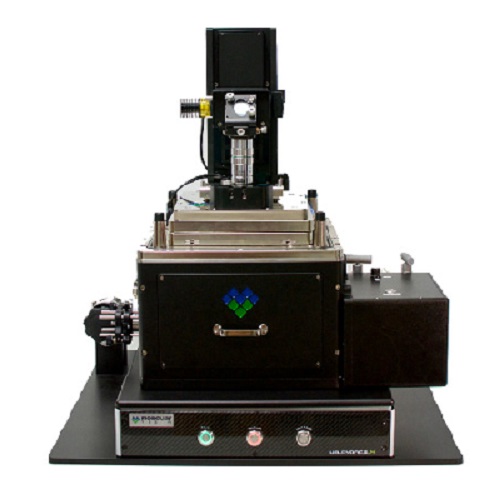
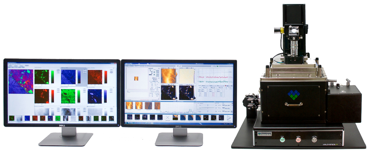
Vista-IR
Vista-IR comes equipped with a mid-IR laser with the widest tuning range and the best beam stability. The same laser can be used for imaging, extremely fast spectrum acquisition, and even for additional techniques such as scattering scanning near-field microscopy (s-SNOM option must be added). Many automated and user-aid features make the tool robust and as easy as using a standard AFM.
Widely Tunable Mid-IR Source
Continuously tunable over 1,000 cm-1 with 1 cm-1 resolution with enough power to generate point spectrum in about ~0.1 sec.
Automated Setup
PiFM setup is simple – the software detects the two cantilever resonances automatically and sets up the laser so that both AFM topography and PiFM images are acquired concurrently.
Simple Alignment
Cantilever is auto-aligned for AFM detection via cantilever alignment chip. IR beam alignment is as simple as taking a 15-second beam profile image (acquired by scanning the parabolic mirror) and clicking the center of the beam profile.
hyPIR ® – Mapping
hyPIR (hyperspectral PiFM IR) mapping consists of PiFM spectrum in each pixel of (n x n) pixel-image. It is like taking a thousand PiFM images and stacking them on top of each other.
High NA (1.45) Inverted Optics
High NA inverted objective lens on 3D piezo scanner for confocal and tip-enhanced optical measurements on transparent substrates.