


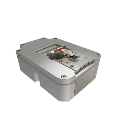
The LiteScope™
provides a wide range of Scanning Probe Microscopy (SPM) imaging modes, which
can be easily used via replaceable probes. Comprehensive sample
analysis including: The LiteScope™ may also be
combined with other SEM accessories: for fabrication of nano/microstructures
and surface modifications. In this combination, the LiteScope™ offers easy and
fast 3D inspection of manufactured structures. The LiteScope™ provides and supports a
wide spectrum of SPM measurement methods and probes.The cornerstone and most
valuable technical feature of its design is the universal probe holder enabling
very easy “Plug & Play” installation of different probes.Imaging modes
CPEM Correlative microscopy is an
approach that benefits from the imaging of the same object by two different
techniques.

LiteScope™ Data LiteScope™ is usually used in high vacuum, but may also
be adapted for ultra high vacuum conditions on
request. Application In situ SEM/AFM characterisation of hybrid
structures made of graphene-veiled gold nanoparticles for bio sensing The
combination of gold nanoparticles and graphene veiling is a novel approach to
fabrication of active substrates suitable for Surface Enhanced Raman
Spectroscopy (SERS). Graphene can serve as a pin-hole passivation layer, which
prevents plasmonic nanostructures from oxidizing. The level of the contact
between Au particles and graphene plays a significant role in the sensitivity
of SERS. Therefore, it is important to know the way a graphene membrane veils a
single nanoparticle or a cluster of such nanoparticles. The distribution of the
nanoparticles under the graphene membrane as well as the surface topography can
be easily determined using CPEM – Correlative Probe and Electron Microscopy™.
While LiteScope™ SPM (AFM) can image the surface of a graphene layer on top of
nanoparticles, SEM can image nanoparticles under the graphene layer. AFM images
show that graphene does not completely wrap the nanoparticles, this results in
reduced SERS effect. Decomposition of annealed solid solution W-Cr
with HfO2particles This
application is focused on the study of microstructure of sample which shows the
decomposition of W-10Cr-1Hf (solid solution W-Cr with HfO2 particles) as a
result of annealing at 1000 °C for 10 hours. The pores are formed around the
hafnium dioxide particles and grow due to the decomposition, also Cr – rich
part (dark lamellae or dots) and W – rich part (bright area between the
lamellae) are constituted. Using CPEM, it is possible to quickly and accurately
distinguish the topographic and the material contrast in SEM images. It is also
possible to easily distinguish the grains of Hf from the pores. It was found
out different etching rate of the sample surface by CPEM technique. AFM
image, SE image (BSE detector) and CPEM image of sample. Study of mesenchymal stem cells on collagen
scaffold Collagen
scaffold was prepared by friendly freeze drying method (Ceitec BUT, RG 2.3).
Then, the scaffold was seeded with mesenchymal stem cells and was
incubated under standard culture conditions for 6 days (Department of
Histology and Embryology, Masaryk University). Finally, the scaffold was
stained with osmium tetroxide and uranyl acetate for better visualization. It
was possible to carry out measurement of prepared cells only due to unique
LiteScope™ and its precise AFM tip navigation by SEM without damaging the probe
in the pores of scaffold. Using CPEM, it was possible to scan the same cell
with both the electron beam and the probe and obtain the images. These images
are then used to obtain topographic information about the cell and its
interaction with the substrate in the meaning of greater spreading of the cell
after the scaffold - better adhesion and a more friendly environment.


